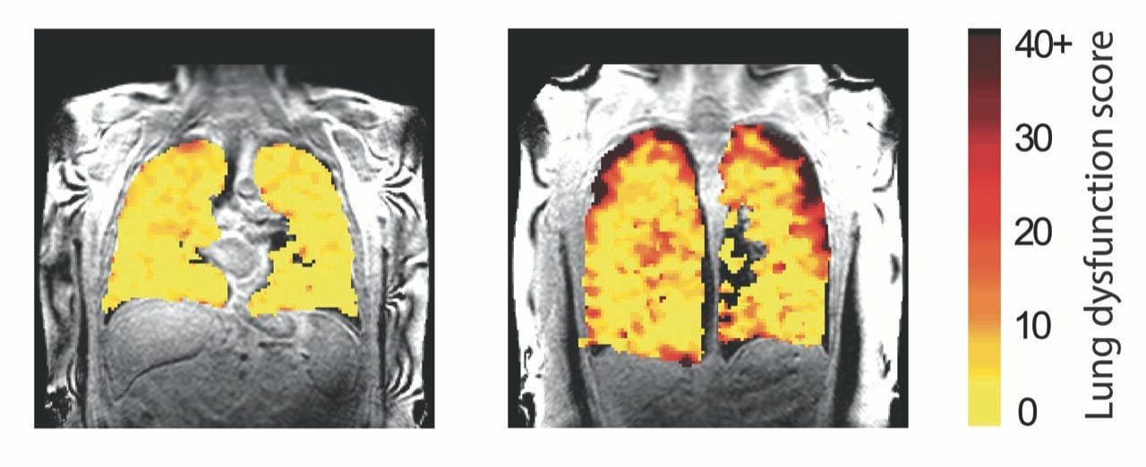
A groundbreaking method for scanning lungs has emerged that could revolutionize the way healthcare professionals monitor lung function and track the effects of treatment in real time. This new scanning technique allows experts to observe how air moves in and out of the lungs, making it possible to monitor the condition of patients with asthma, chronic obstructive pulmonary disease (COPD), and even those who have received lung transplants. This innovative technology holds the potential to enable medics to detect early signs of lung function decline, which could significantly improve patient outcomes and inform the management of respiratory conditions.
The method, developed by researchers at Newcastle University in the UK, utilizes a special gas called perfluoropropane that can be safely inhaled and exhaled by patients. When this gas is inhaled, it can be visualized using magnetic resonance imaging (MRI). This allows clinicians to see precisely where the gas has reached within the lungs, providing a detailed view of lung function that wasn’t possible with traditional imaging techniques. The ability to track air movement within the lungs gives researchers and healthcare professionals a unique, non-invasive tool for assessing and monitoring the lungs’ performance, particularly in patients with chronic lung diseases and those who have undergone lung transplants.
The team, led by Professor Pete Thelwall, who is a professor of Magnetic Resonance Physics and the director of the Center for In Vivo Imaging at Newcastle University, conducted studies to explore how this new scanning technique works in patients with asthma, COPD, and those who have received a lung transplant. Two complementary papers detailing their research were published in Radiology and JHLT Open. These studies demonstrate how the scan provides a detailed, real-time analysis of lung function, giving clinicians valuable insights into areas of the lungs that may be underperforming or not receiving adequate ventilation.
The perfluoropropane gas used in the scan provides a clear picture of how air is distributed throughout the lungs during normal breathing. In patients with respiratory conditions like asthma and COPD, this scan can identify regions of the lung where air does not reach effectively, a feature that is critical for assessing disease progression and treatment efficacy. For example, when patients use their asthma medication, the scan reveals which areas of the lung are better able to move air in and out, allowing doctors to track the effectiveness of the treatment. This dynamic monitoring method can lead to better, more personalized management of respiratory conditions, as it shows exactly which parts of the lung improve or worsen over time.
In the case of patients with asthma or COPD, the team demonstrated how this new scanning method could be used to quantify improvements in ventilation following treatment with bronchodilators, such as salbutamol. In their study, the team showed that this technique could be valuable in clinical trials of new lung disease treatments by providing a sensitive and real-time measure of lung function improvement. This is a significant advancement in how we approach the treatment of respiratory diseases, as it allows for much more precise tracking of patient progress.
In a further development of the scanning technique, the team conducted a study involving lung transplant recipients, published in JHLT Open. Lung transplant patients are often at risk of chronic lung rejection, a condition in which the immune system attacks the transplanted lung tissue. This is a common issue among transplant recipients, and it can significantly impact lung function. By using the new scanning method, the research team was able to track air movement in transplanted lungs, providing valuable insight into how well the transplanted lungs are functioning over time.
In transplant recipients, the scan revealed how air moved differently in those with chronic rejection compared to those with normal lung function. Specifically, patients with chronic rejection showed poorer airflow to the edges of the lungs, which is characteristic of chronic lung allograft dysfunction (CLAD). CLAD is a common complication after lung transplants, and early detection is critical to managing the condition before irreversible damage occurs. By identifying changes in lung function at an earlier stage than traditional diagnostic tests, this new method of scanning could enable clinicians to initiate treatment sooner and protect the transplanted lungs from further damage.
Professor Andrew Fisher, a co-author of the study and a professor of respiratory transplant medicine at Newcastle Hospitals NHS Foundation Trust and Newcastle University, emphasized the potential of this scanning technique in improving the care of lung transplant patients. He explained, “We hope this new type of scan might allow us to see changes in the transplant lungs earlier and before signs of damage are present in the usual blowing tests. This would allow any treatment to be started earlier and help protect the transplanted lungs from further damage.”
The research team also highlighted that this new scanning method has broader implications for the future of lung disease management, including its potential to be used in routine clinical practice. By providing a sensitive and detailed measure of lung function, the scan could help doctors detect early changes in lung health, even before symptoms become apparent or before traditional diagnostic tests show abnormalities. This could be particularly useful in monitoring patients with chronic lung diseases like COPD, as well as in assessing the long-term health of lung transplant recipients.
The ability to detect subtle changes in lung function before they become clinically significant is a major breakthrough in the management of lung disease. Early intervention is key to preventing further damage and improving the overall prognosis for patients. This technology has the potential to transform the way we monitor and treat respiratory conditions, making it possible to deliver more personalized and effective care.
Overall, the development of this new MRI scanning technique represents a significant step forward in the diagnosis and treatment of lung diseases, particularly for patients with asthma, COPD, and those who have undergone lung transplants. By providing real-time insights into lung function, this method offers a more dynamic and accurate picture of a patient’s respiratory health. As the technology continues to evolve, it is likely that it will become an essential tool in the management of lung diseases, enabling clinicians to provide better care and ultimately improving the quality of life for patients worldwide.
References: Pippard BJ, et al, Assessing Lung Ventilation and Bronchodilator Response in Asthma and Chronic Obstructive Pulmonary Disease with Fluorine 19 MRI, Radiology (2024). DOI: 10.1148/radiol.240949
Mary A. Neal et al, Dynamic 19F-MRI of pulmonary ventilation in lung transplant recipients with and without chronic lung allograft dysfunction, JHLT Open (2024). DOI: 10.1016/j.jhlto.2024.100167
Think this is important? Spread the knowledge! Share now.
