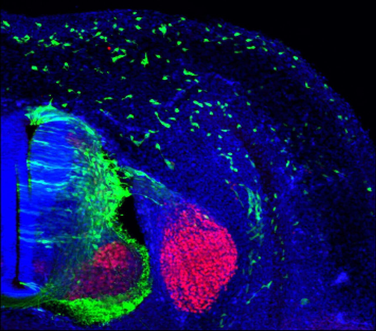
An international team of researchers has made a groundbreaking discovery about the genetic mechanisms that regulate the formation and migration of cranial neural crest cells, critical to the development of facial structures during embryonic growth. Their findings, recently published in The American Journal of Human Genetics, delve into the interplay between specific genes and their roles in a developmental process known as epithelial-to-mesenchymal transition (EMT), offering new insights into congenital disorders and their underlying genetic causes.
At the core of this research lies the discovery of how the ZIC2 gene, working alongside the ARID1A-BAF complex, orchestrates the process of EMT. This fundamental mechanism allows cells to undergo shape changes and migrate to their destined locations within the developing embryo, forming key tissues and organs, including the facial architecture. Disruption in this process has been implicated in various congenital abnormalities, making the study pivotal in understanding developmental disorders that affect craniofacial structures.
The collaborative study was co-led by Dr. Eloísa Herrera, head of the Generation and Regeneration of Bilateral Neural Circuits laboratory at the Institute of Neurosciences (a joint initiative by the Spanish National Research Council and Miguel Hernández University of Elche), and Dr. Marco Trizzino, leader of a research group specializing in human stem cells at Imperial College London. Together, the international team employed cutting-edge techniques and interdisciplinary approaches to uncover how genetic anomalies in the ARID1A-ZIC2 pathway impact neural crest cell development.
The neural crest, a unique group of cells in vertebrate embryos, is a critical contributor to the formation of craniofacial features, peripheral nerves, and certain cardiac structures. During EMT, neural crest cells transform from static epithelial cells into highly mobile mesenchymal cells, enabling them to travel to their designated locations. This ability to migrate and differentiate is essential for forming various tissues, including the cartilage, bone, and connective tissue that make up the face. Understanding how this transformation is regulated at the genetic level is vital for uncovering the causes of craniofacial malformations.
To better comprehend these mechanisms, the researchers studied cells derived from patients with Coffin-Siris Syndrome (CSS). This rare genetic disorder, caused by insufficient ARID1A gene function, is characterized by diverse abnormalities such as craniofacial defects, limb malformations, and intellectual disabilities. By analyzing these cells, the team could observe how mutations in the ARID1A gene disrupt the genetic programming involved in EMT and, consequently, the migration of neural crest cells.
Using advanced RNA sequencing (RNA-seq) and chromatin immunoprecipitation sequencing (ChIP-seq), the scientists mapped the genetic programs regulated by the ARID1A-ZIC2 axis. These techniques allowed them to identify the genes directly influenced by this molecular pathway and understand how their activity impacts craniofacial development. Their results revealed that ARID1A plays a critical regulatory role by enabling ZIC2 to activate EMT-related genes at specific genomic sites. Without proper ARID1A function, ZIC2 cannot effectively occupy these key regions, leading to a failure in neural crest migration and the manifestation of craniofacial abnormalities.
To validate their findings, the team employed in vivo models using mice and chicken embryos. These models provided valuable insights into how the ARID1A-ZIC2 pathway functions within a living organism and what happens when its activity is disrupted. By examining these animal embryos, the researchers demonstrated that ZIC2 is prominently expressed in premigratory neural crest cells, marking the critical stage just before these cells begin their migration. Furthermore, the absence of ARID1A was shown to lead to significant defects in craniofacial development, confirming the essential role of this genetic pathway.
Dr. Herrera emphasized the importance of understanding these mechanisms, stating, “We were able to observe the precise timing and spatial expression of ZIC2 within neural crest cells, which provided a clearer picture of its role in regulating migration. Disrupting this process not only affects craniofacial structures but also sheds light on the broader implications of ARID1A-related genetic disorders.”
The study underscores the significance of ARID1A in regulating the genetic blueprint for EMT and highlights ZIC2 as a crucial player in this process. By uncovering how these two components interact, the research team has opened new avenues for exploring potential therapies. Targeted treatments could potentially address craniofacial malformations and other developmental disorders stemming from genetic disruptions.
Beyond its implications for developmental biology, the research also has broader clinical relevance. Congenital craniofacial malformations, such as cleft lip, cleft palate, and craniosynostosis, are among the most common birth defects globally, often resulting in significant medical, emotional, and social challenges for affected individuals. Understanding the genetic underpinnings of these conditions is a vital step toward improving diagnostic tools, preventative strategies, and therapeutic interventions.
Dr. Trizzino highlighted the translational potential of this discovery, saying, “Our findings provide a molecular framework for understanding congenital diseases associated with neural crest defects. By targeting the ARID1A-ZIC2 pathway, we may be able to develop interventions that restore normal cellular function and prevent or mitigate these conditions.”
The study also exemplifies the importance of international collaboration in advancing scientific research. By combining expertise from different institutions and leveraging diverse experimental systems, the team was able to piece together a complex puzzle that sheds light on a previously elusive aspect of human development. Their work not only deepens our understanding of how facial structures are formed but also illustrates the power of collaborative efforts in addressing fundamental questions in biology.
Reference: Samantha M. Barnada et al, ARID1A-BAF coordinates ZIC2 genomic occupancy for epithelial-to-mesenchymal transition in cranial neural crest specification, The American Journal of Human Genetics (2024). DOI: 10.1016/j.ajhg.2024.07.022


