Researchers at the University of Adelaide have made an exciting breakthrough in the world of life sciences by using quantum technology to image embryos for the first time. This pioneering research marks a significant leap in our ability to observe and understand the delicate processes of life at a microscopic level, with the potential to revolutionize fields like reproductive health and developmental biology.
The research was conducted by the University’s Center of Light for Life, a hub of academic excellence focused on exploring how light can be harnessed to better understand biological systems. Led by Professor Kishan Dholakia, a leading figure in optics and photonics, the team utilized advanced imaging technology to capture images of living embryos. What makes this study unique is the use of specialized cameras designed for quantum measurements—technology that is typically employed in high-precision fields like physics and astronomy.
The Role of Photons in Embryo Imaging
At the heart of this research is the ability to detect individual packets of light energy, known as photons. These ultra-sensitive cameras are designed to count the photons that interact with the specimen at each pixel, providing an exceptionally detailed image. The significance of this technology lies in its ability to observe biological processes in their natural state, without damaging the samples—a common problem with conventional imaging methods.
Professor Dholakia highlighted the importance of minimizing potential harm to the samples being observed, particularly when studying live cells and embryos. “Damage from illumination is a real concern,” he explained. “The more light we shine on these cells, the more likely we are to affect their behavior or development. Using the lowest possible light level, combined with sensitive cameras, allows us to understand biology without compromising the integrity of the cells.”
This careful balance between capturing detailed imagery and protecting the specimen is one of the key innovations in this research. Traditional imaging methods often use bright lights or lasers to capture images, but these intense light sources can be harmful to living cells. By using the least amount of light necessary for the task, and pairing it with ultra-sensitive cameras, the team has managed to open new doors in biological imaging, preserving the natural state of the cells while still gaining incredibly detailed insights.
Breaking Boundaries in Imaging Technology
The team’s innovative approach was recently published in APL Photonics, where they detailed the use of these cutting-edge cameras to image embryos as part of a pre-clinical trial. This trial forms a crucial step in understanding how these imaging techniques could be applied in clinical practices, such as in vitro fertilization (IVF) and early-stage embryo development. As Professor Dholakia points out, “These samples are living, developing specimens that serve as a foundation for studies supporting advancements in clinical IVF.” This could mean more effective and safer treatments for people undergoing IVF, where understanding embryo development is crucial for increasing the success rate of pregnancies.
The use of quantum cameras in life sciences is still in its infancy, but it holds vast potential for the future. Lead author and Ph.D. student Zane Peterkovic noted how this project bridged the gap between advanced physics and biology: “Digital camera technology has advanced to the point where fundamental physics concepts like quantum mechanics become important and relevant.” In particular, quantum cameras allow researchers to capture weak signals emitted by natural compounds in cells. These compounds, when illuminated, can provide a wealth of information about the biological processes taking place—but the signals they emit are often too faint for traditional cameras to detect effectively. By using quantum cameras that can count individual photons, the team is able to capture these weak signals and extract meaningful data from them.
Overcoming Challenges and Enhancing Image Quality
One of the most challenging aspects of this project was developing a method to fairly compare image quality across different types of cameras. Traditional imaging techniques often involve taking a snapshot and analyzing the resulting image. However, with quantum cameras, the process is more complex due to the sensitivity and the nature of the light being captured.
A significant portion of the research focused on fine-tuning the imaging process to get the most out of these specialized cameras. Peterkovic explained, “A large part of the project involved developing a method to fairly compare the image quality across different cameras.” This involved not only using advanced imaging hardware but also employing specialized software and computational methods to enhance the final results.
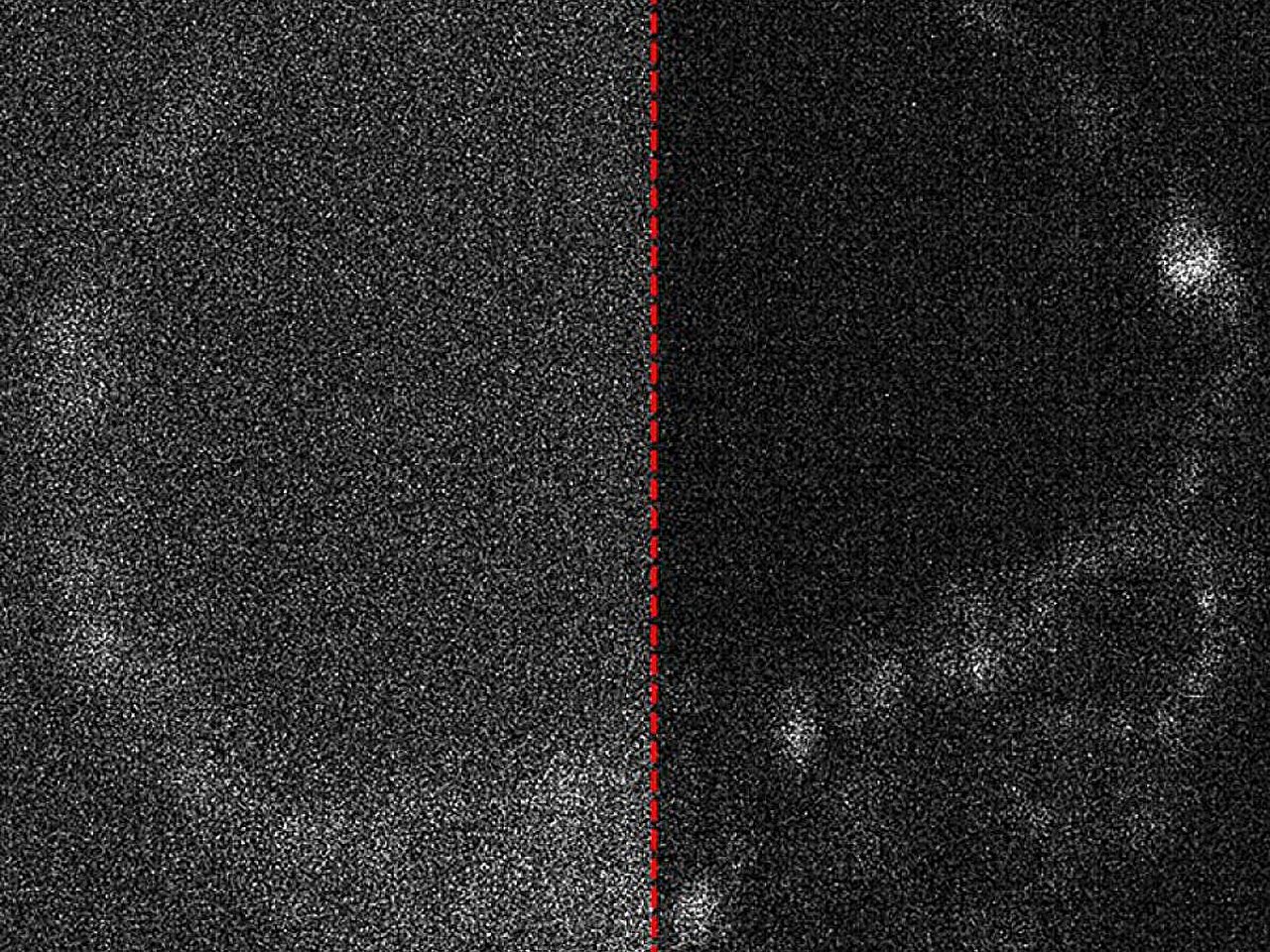
To combat the problem of weak signals and static in the images, the team explored the use of artificial intelligence (AI) to “clean up” the images. The challenge lies in the fact that quantum cameras struggle to capture enough light, leading to “noise” in the images—static or interference that can obscure important details. AI-powered algorithms can help filter out this noise, making the images clearer and more accurate. Peterkovic and his colleagues developed a method for applying AI to remove noise, ensuring that the final images were as sharp and informative as possible.
“We even explored how AI can be used to remove noise from the captured images,” said Peterkovic. “These steps go beyond just putting the camera in the microscope to take pictures.” The combination of quantum cameras and AI is paving the way for the next generation of imaging technology—one that can capture the unseen details of biology in a way that was previously thought impossible.
The Future of Quantum Imaging in Life Sciences
Looking ahead, the research team is excited about the potential applications of quantum imaging technology. Future directions for this work include extending the technology into the realm of quantum imaging, where the quantum states of light could be used to extract even more detailed information about the biological samples. This could open up new possibilities for studying the structure of living cells, tracking the movement of molecules, and even observing chemical reactions in real time.
Quantum imaging could also have far-reaching implications in medical diagnostics. By observing biological samples at a much finer scale than currently possible, it may be possible to detect early signs of diseases like cancer or Alzheimer’s, where subtle changes in cell behavior can be indicators of developing conditions. Quantum cameras could enable doctors to make more accurate diagnoses and offer more personalized treatment plans based on detailed imaging of a patient’s cells or tissues.
The team also sees potential applications for this technology in fields like neuroscience, where understanding the behavior of neurons and brain cells is crucial for advancing treatments for mental health disorders. The ability to study live, developing brain cells with minimal disruption could lead to breakthroughs in understanding conditions such as autism, schizophrenia, and depression.
Conclusion
The research conducted by the University of Adelaide’s Center of Light for Life represents a major step forward in the application of quantum technology to the life sciences. By using cameras designed for quantum measurements, the team has unlocked the ability to image embryos and living cells with unprecedented detail, all while minimizing potential damage from light exposure. This work opens up new possibilities for understanding biology, from the earliest stages of life to complex cellular processes, and has the potential to revolutionize medical diagnostics and treatments in the future.
The combination of advanced imaging technology, quantum mechanics, and AI offers an exciting glimpse into the future of biological research. With further developments and applications, quantum imaging may become a key tool in unraveling the mysteries of life itself, paving the way for a new era of scientific discovery.
Reference: Zane Peterkovic et al, Optimizing image capture for low-light widefield quantitative fluorescence microscopy, APL Photonics (2025). DOI: 10.1063/5.0245239
Think this is important? Spread the knowledge! Share now.