Over the course of their lives, humans and other animals learn to avoid situations and stimuli that are dangerous or perceived as threatening. This ability to recognize and respond to potential threats is crucial for survival and is deeply embedded in the brain’s neural circuitry. While past neuroscience studies have established that the medial prefrontal cortex (mPFC) plays a key role in learning and decision-making, its specific involvement in the gradual development of threat responses has remained largely unexplored.
Recent research from the University of California, Los Angeles (UCLA) has provided new insights into how the brain’s neural connections develop over time and influence changes in threat responses. The study, published in Nature Neuroscience, revealed that distinct transitions during brain development impact how the mPFC interacts with two key regions involved in emotional learning: the nucleus accumbens (NAc) and the basolateral amygdala (BLA). These findings could have profound implications for understanding mental health disorders, particularly those linked to early life stress and trauma.
The Neural Basis of Threat Learning
The ability to recognize and respond to threats is controlled by a complex network of brain regions that process fear, decision-making, and emotional learning. The limbic system, often referred to as the brain’s emotional center, plays a crucial role in these processes. The medial prefrontal cortex (mPFC) is a particularly important region that governs top-down control over threat-related behaviors, meaning it helps regulate responses to fearful situations based on past experiences.
Dysfunction in these neural circuits has been linked to anxiety, mood disorders, and substance use disorders, many of which emerge during adolescence. “Our study began when we noticed a key gap in our understanding of how neural circuits in the limbic system—the brain’s emotion centers—develop,” said Laura DeNardo, senior author of the study.
Past research has consistently shown that adverse experiences during childhood, such as chronic stress or trauma, can significantly increase the risk of developing mental health disorders later in life. However, most studies examining the connection between early life adversity and mental health were conducted on adults, leaving a gap in understanding how brain development is affected at a cellular level.
“To understand how risk factors like early life stress influence mental health, we require a basic understanding of the developmental sequences that shape the maturation of the limbic system,” DeNardo explained. “This knowledge will provide a foundation for understanding how different forms of early life adversity—varying in nature, severity, and timing—alter developmental trajectories in the limbic system, potentially leading to distinct mental health disorders.”
Modeling Risk-Taking and Threat Avoidance in Mice
To explore how the brain develops its ability to recognize and avoid threats, DeNardo and her team conducted experiments using mice as a model organism. They designed a new behavioral assay (experimental task) to study risk-taking behavior in adolescent mice.
In this experiment, the mice were trained to avoid a signaled threat by stepping onto a safety platform when presented with a danger cue. Interestingly, juvenile and adolescent mice exhibited lower levels of threat avoidance compared to adults, mirroring patterns observed in human adolescents, who often take more risks compared to children or adults.
This behavior aligns with previous findings suggesting that the adolescent brain has not yet fully developed the neural mechanisms required for optimal threat assessment and decision-making. This phase of development might be an evolutionary adaptation, allowing younger individuals to explore and take risks that could lead to greater opportunities, despite the potential dangers.
Tracking and Manipulating Brain Activity
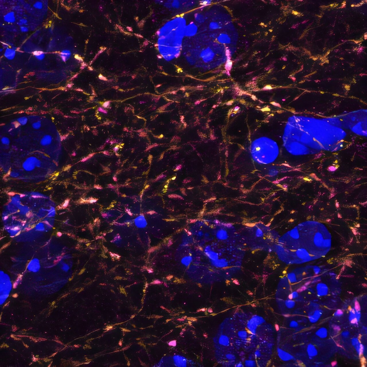
To understand how neural circuits contributed to these behaviors, the researchers used viral expression techniques to introduce molecules into the mice’s brains. These molecules enabled them to track and manipulate neural activity as the animals engaged in the threat avoidance task.
They employed a technique called fiber photometry, which allows scientists to measure neural activity in living animals in real time. This method was used to examine activity in the three key brain regions involved in emotional regulation and fear-based learning:
- Medial prefrontal cortex (mPFC) – Governs cognitive control over emotions and decision-making.
- Nucleus accumbens (NAc) – Involved in motivation, reward, and action selection.
- Basolateral amygdala (BLA) – Processes fear and emotional memories.
The data revealed that in adult mice, the mPFC showed stronger neural responses to both threat-predictive cues and safe locations. In contrast, juvenile and adolescent mice had weaker mPFC activity, suggesting that this brain region is still developing and plays a limited role in early life threat responses.
However, activity in the NAc and BLA was more consistent across different age groups, indicating that these regions might develop faster than the mPFC. These findings support the idea that mPFC-based control over emotional learning is a gradual process that continues into adulthood.
Using Optogenetics to Control Brain Circuits
To further investigate these developmental differences, the researchers used optogenetics, a technique that allows precise control of neurons using light-sensitive proteins. By selectively stimulating or suppressing neural activity in mPFC pathways, they observed significant changes in the mice’s behavior.
Their experiments showed that different mPFC pathways influenced threat avoidance behavior in distinct ways, depending on the animal’s age. This suggests that the function of these pathways evolves over time, shaping how individuals learn to avoid threats at different life stages.
Additionally, they combined optogenetics with immediate early gene staining, a technique that marks neurons that were recently active. This approach allowed them to identify specific cell types in the NAc and BLA that interact with mPFC circuits during different developmental stages.
Revisiting Theories of Adolescent Brain Development
One widely accepted theory of adolescent brain development suggests that weak prefrontal control over emotional centers like the NAc and BLA leads to riskier behaviors in younger individuals. As the mPFC matures, stronger top-down control emerges, allowing for better regulation of emotions and threat responses.
However, DeNardo’s study challenges this gradual development model, showing that while some synaptic changes occur progressively, others happen in sudden shifts. For example, while mPFC-NAc connections gradually strengthen over time, mPFC-BLA connections remain stable until adulthood, when they undergo rapid changes.
This finding suggests that different mPFC pathways develop at different rates, each serving unique functions at different life stages. Rather than a uniform increase in prefrontal control over emotional centers, the development of these neural circuits is more dynamic and punctuated.
Implications for Mental Health and Future Research
This research provides new insights into how the brain matures and adapts its ability to process and avoid threats. These findings could have important implications for understanding mental health conditions like anxiety, PTSD, and substance use disorders, which are often linked to early life stress and trauma.
By identifying critical periods of neural development, researchers may be able to design interventions that prevent or mitigate the effects of early life adversity. Understanding how different forms of childhood trauma—whether chronic stress, neglect, or traumatic events—alter brain development could lead to more effective treatments for mental health disorders.
DeNardo and her team are currently investigating how early life stress affects the mPFC-BLA pathway and influences threat avoidance behavior in adolescence. This future research could provide crucial insights into why some individuals are more vulnerable to anxiety and PTSD than others.
Conclusion
The UCLA study reveals that the maturation of threat avoidance behaviors is not a simple, linear process. Instead, different brain regions and pathways develop at different rates, with sudden shifts occurring at key developmental stages. These findings challenge traditional models of adolescent brain development and highlight the complex, dynamic nature of neural circuit maturation.
As neuroscience continues to uncover the mechanisms behind emotional learning and fear regulation, this knowledge may lead to better strategies for treating mental health disorders. By understanding how early experiences shape the brain, researchers may ultimately help develop more effective interventions to support mental well-being across the lifespan.
Reference: Cassandra B. Klune et al, Developmentally distinct architectures in top–down pathways controlling threat avoidance, Nature Neuroscience (2025). DOI: 10.1038/s41593-025-01890-w