Interleukin-1 (IL-1) is a potent cytokine, or signaling molecule, that plays a critical role in regulating immune responses, inflammation, and maintaining homeostasis in both healthy and diseased states. Although IL-1 is most commonly associated with inflammatory responses in the body, recent research has highlighted its significant involvement in the brain, contributing to both the regulation of normal brain functions and the exacerbation of neuroinflammation in various disorders, particularly those related to stress, mood, and cognition.
IL-1 exists in two main forms: IL-1α and IL-1β, both of which share similar receptor binding properties and exert key roles in activating inflammatory pathways in the body and the central nervous system (CNS). Understanding how IL-1 functions within the brain under normal and diseased conditions is critical for uncovering novel therapeutic avenues for neurological and psychiatric disorders.
The Dual Nature of IL-1 in the Brain
Under typical conditions where no inflammation is present, IL-1 plays an essential role in regulating several brain functions. It influences the production of hormones, helps to modulate sleep patterns, and enhances cognitive abilities such as memory and learning. Despite these beneficial functions, elevated levels of IL-1 within the brain are closely associated with various forms of brain inflammation, which can lead to adverse effects on both physical and mental health.
Neuroinflammation, the chronic activation of immune cells in the brain, is implicated in many neurodegenerative conditions, including Alzheimer’s disease, Parkinson’s disease, and multiple sclerosis, as well as mental health conditions such as depression and anxiety. In such diseases, excess IL-1 can exacerbate the inflammatory response, worsening brain tissue damage and promoting the onset or progression of illness. Research has shown that high IL-1 levels in the brain disrupt the body’s stress response, induce sickness-like behaviors (such as fatigue and lack of motivation), and potentially allow immune cells from the body to enter the brain. These responses can trigger a cascade of damage, with the support cells in the brain, such as glial cells, producing molecules that further damage neuronal networks. Additionally, excessive IL-1 can lead to cognitive impairments, particularly related to mood disorders and the deterioration of memory and thinking.
The Role of IL-1R1: A “Doorbell” for Immune Responses
IL-1 exerts its influence on brain function through its receptor, Interleukin-1 Receptor Type 1 (IL-1R1). This receptor can be likened to a “doorbell” on the surface of cells. When there is an infection, injury, or inflammation in the body, IL-1 binds to IL-1R1, triggering a signaling cascade that activates immune responses. While IL-1R1’s role in immune cells is well-established — namely in the initiation of inflammatory responses — its effects on neurons (the brain’s nerve cells) have been a topic of considerable research interest.
Despite the widespread assumption that IL-1R1 signaling in neurons would induce inflammation, recent studies suggest a more nuanced role for IL-1R1 in brain function. Specifically, neurons that express IL-1R1 do not appear to induce traditional inflammatory responses. Instead, these neurons are believed to integrate immune signals into neural activity, potentially influencing various cognitive and emotional functions. Researchers continue to explore exactly how IL-1R1 signaling in neurons controls or modifies normal brain processes, such as sensory perception, mood regulation, and memory.
New Insights into Neuronal IL-1R1 Expression
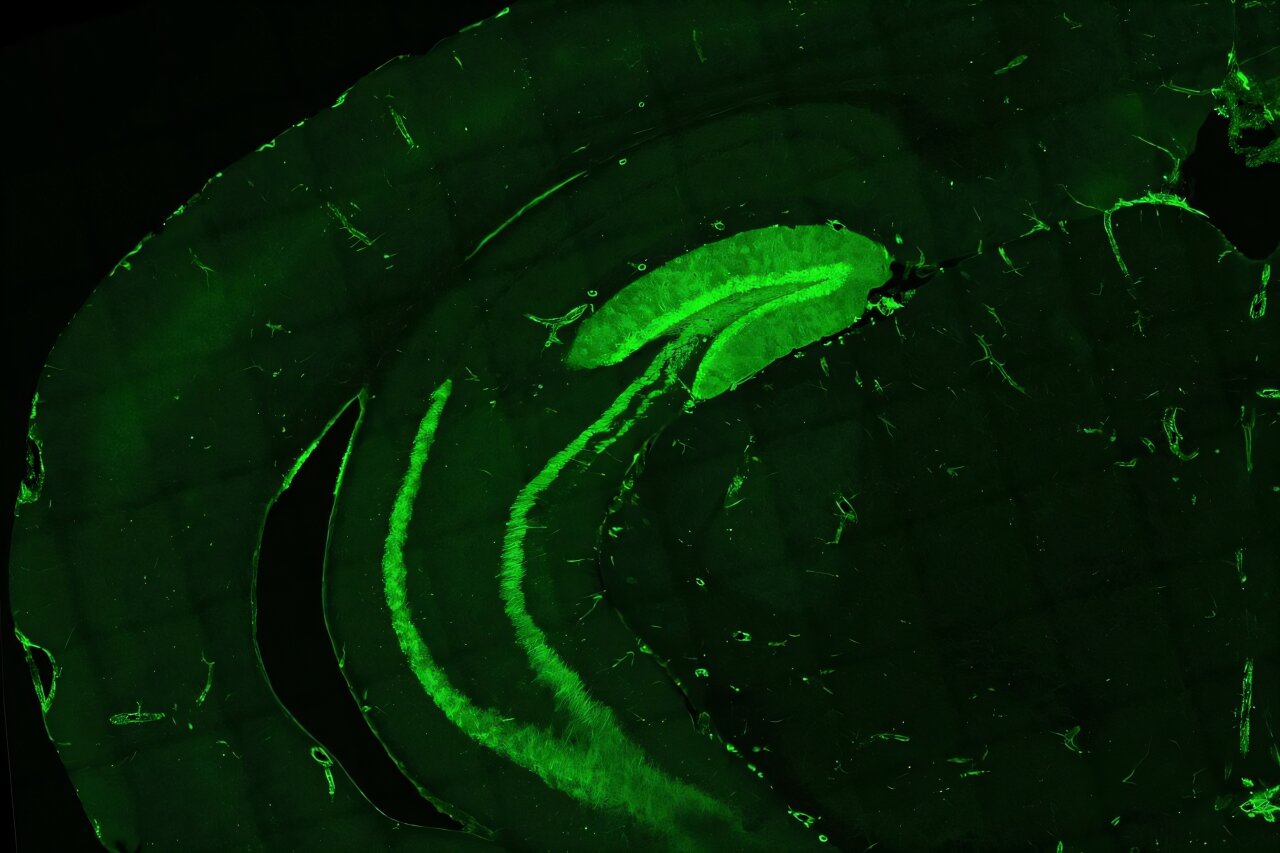
A recent study from Florida Atlantic University provides the most comprehensive mapping of neuronal IL-1R1 expression in the mouse brain to date. Published in the Journal of Neuroinflammation, this research marks a significant advancement in our understanding of IL-1 signaling in the brain, filling in gaps from previous research. Prior work has suggested that IL-1 signaling in neurons could affect behaviors such as sickness behavior, anxiety, and changes in sleep, but the exact neural circuits involved were poorly understood. The FAU study aimed to identify specific neuronal populations and neurotransmitter systems that are likely responsible for these effects.
Using genetically modified mice, the researchers employed a novel cell tagging approach to pinpoint neurons that express IL-1R1. This allowed the team to identify key brain regions and cellular circuits involved in mediating IL-1R1-related functions. The findings provide new insights into how inflammation in the brain impacts cognitive, sensory, and emotional processes.
Key Findings and Implications for Brain Disorders
The study’s most significant revelation was that IL-1R1 expression is most prominent in brain regions critical for sensory processing, mood regulation, and memory, with particular emphasis on the somatosensory cortex and glutamatergic (excitatory) systems. The somatosensory cortex, for example, processes sensory information from the body, while glutamatergic neurons are involved in signaling pathways that promote learning and memory.
Researchers found that many of these IL-1R1-expressing neurons use glutamate as their primary neurotransmitter. Additionally, the study discovered that neurons in these regions also utilize serotonin, a neurotransmitter critical for mood regulation. This dual neurotransmitter signaling suggests that neuronal IL-1R1 is involved in circuits controlling a wide range of behavioral and cognitive functions, including emotional responses and cognitive flexibility. Importantly, IL-1R1 expression was not only found in areas traditionally associated with cognitive and emotional processing but also in sensory processing centers, such as the thalamus and various sensory cortical regions.
The new findings reveal that IL-1R1 expression is closely tied to specific gene pathways involved in synapse organization — the physical connections between neurons — but does not cause the typical inflammatory response commonly associated with IL-1 activation in other cell types. This indicates that IL-1R1 might play a critical role in shaping neural circuits and maintaining their function without triggering full-fledged inflammation, which was a previously unexpected discovery. This insight broadens our understanding of how immune signaling could influence brain structure and function without directly inducing neuroinflammation.
According to Dr. Ning Quan, the senior author of the study and a researcher at FAU’s Schmidt College of Medicine, this work illuminates how certain neurons may integrate immune signals and how this integration could help explain the connections between neuroinflammation and disorders such as depression, anxiety, and cognitive dysfunction. Dr. Quan suggests that IL-1R1 signaling influences the neural circuits that govern behavior, potentially opening the door for new therapeutic approaches to mental health disorders linked to chronic inflammation.
Implications for Treating Inflammatory Brain Disorders
The findings of this study have important implications for understanding how brain inflammation contributes to a range of disorders and could lead to new treatment strategies for conditions like chronic stress, depression, and anxiety. As Dan Nemeth, the first author of the paper, notes, the study offers new insights into the fundamental biological mechanisms that govern not only mood and memory but also sensory processing — an area that had previously been understudied in this context.
By identifying the precise brain regions and neural circuits that mediate IL-1R1 signaling, this study paves the way for more targeted interventions in neuroinflammatory disorders. As Dr. Randy D. Blakely, a co-author and the executive director of the Stiles-Nicholson Brain Institute, suggests, the study opens the door to potential therapies that could modify or block IL-1 signaling pathways in the brain to mitigate the behavioral and cognitive impairments seen in mood and anxiety disorders.
Conclusion
The findings from this groundbreaking study provide critical insights into how IL-1R1 signaling influences brain function, particularly in relation to sensory processing, mood regulation, and memory. By offering the most comprehensive mapping of neuronal IL-1R1 expression to date, researchers have refined our understanding of how immune signaling is linked to the neural circuits that underpin behavior. These results could inform future research and therapeutic approaches for treating inflammatory-based brain disorders, bringing us one step closer to addressing the complex relationship between brain inflammation and psychiatric conditions.
Reference: Daniel P. Nemeth et al, Localization of brain neuronal IL-1R1 reveals specific neural circuitries responsive to immune signaling, Journal of Neuroinflammation (2024). DOI: 10.1186/s12974-024-03287-1
